As the gold standard for diagnosing many heart diseases, a resting electrocardiogram (ECG) may be the single most crucial thing you do all day for your patients.
It is important that everyone involved in your ECG workflow knows and sticks to the basics. When recording an ECG, electrical noise could be caused by leadwire connections that aren’t fully compatible with the current system.
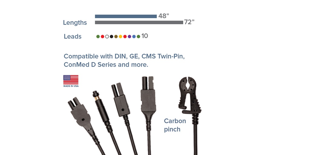
Your monitoring tools would be incomplete without a set of high-quality radiolucent ECG leadwires. When ECG leads are radiolucent, they do not need to be moved or replaced during the fluoroscopy procedures, which speeds up the overall procedure.
Most importantly, radiolucent ECG wires are made with materials that are transparent to X-rays and do not interfere with the view of the procedural area when X-rays are utilized. This allows clinicians to monitor heart function while performing invasive procedures, as radiolucent ECG wires will not interfere with radiographs. This is why it’s best to use high-quality radiolucent lead wires for procedures like stenting and ablation therapy.
The Precise Art of Using Leadwires to Record an Electrocardiogram
Electrocardiography, also called an ECG or EKG, is the study of the electrical activity of the heart. Electrodes are placed on the skin while the myocardium contracts and relaxes to record the electrical activity of the heart. ECG/EKG data is recorded so that it can be analyzed later for heart rate variability, waveform shape, arrhythmia, and other similar conditions.
But it can be hard to get signals that are free of noise, especially when doing a 12-lead ECG. Setting up the equipment to record the data, making sure the subject is comfortable, and placing the electrodes on the subject’s torso and limbs in the right places are just a few of the many things that must be done right to get results that can be correctly interpreted.
Ensure Your Patient Is Well-Prepped
The precision of your ECG reading depends on the patient’s readiness. For an accurate ECG reading, the patient’s skin needs to be cleaned and shaved before the electrodes are put on. Electrode placement is a crucial part of every ECG procedure.
With alcohol-soaked cotton swabs, carefully wipe the chest area to get it ready for the electrodes. This takes away the chance that any oil on your skin could change how accurate your ECG or EKG readings are.
After the skin has been cleaned, the electrode sites can be located and marked.
Using Radiolucent ECG Leadwires for Accurate Precordial Electrode Placement
Six precordial electrodes are initially placed on the patient’s chest. To do this, you need to find the notch in the patient’s sternum. It’s a tiny horizontal ridge that can be felt around 1.5 inches (3.8 cm) down from the top of the sternum. Starting from this point, you can position the electrodes.
Locate V1 and V2 first while trying to determine the proper chest lead location. Because all other chest leads are positioned with respect to V1 and V2, getting these two leads right is crucial.
To locate the fourth intercostal space, begin at the second rib and work your way along the right side of the chest wall. On the right side, V1 is situated close to the sternal border. Feel for the fourth intercostal gap on the left side of the chest, beginning at the second rib. The left sternal border is where V2 should be implanted.
It is very important to find the exact VI position (the fourth intercostal gap) because it is the pivot point for the rest of the electrodes.
Taking biosignal recordings in the Cath labs might be challenging due to the high background noise levels. The radiolucent ECG leadwires from LifeSync not only attach quickly to your monitoring devices but also have low noise levels, which are essential for ECG recordings to achieve good signal quality. This lets you keep an eye on how a patient’s heart is working and catch potentially fatal problems early. To learn more about LifeSync’s radiolucent ECG leadwires, go to rochestersuperstore.com right now.